Confocal laser microscopy is an experimental technique providing with high-resolution
in-focus optical images, which are acquired in raster-mode (i.e. point-by-point) and reconstructed
with a computer.
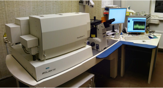
Our laboratory is equipped with modern 3D scanning confocal microscope with spectrometer "Nanofinder-S",
which is used to perform studies with submicrometer resolution (about 250-500 nm) of solid bulk materials,
thin films, nanopowders, solutions, etc.
The system allows to conduct simultaneous and multifunctional analysis by optical and confocal
microscopy; Raman and luminescence spectroscopy; 0D, 1D, 2D and 3D high-speed imaging with spectroscopy.
REFERENCES
A. Kuzmin, Introduction to Nanofinder-S.
A. Kuzmin,
Modernie optiskie mikroskopi (in Latvian).
A. Kuzmin, R. Kalendarev, A. Kursitis, J. Purans,
Confocal Spectromicroscopy of Micro and Nano-Structured Materials.
(Presentation at the 2nd Latvian Conference Functional Materials and Nanotechnologies, Riga, March 27-28, 2006.
A. Kuzmin, R. Kalendarev, A. Kursitis, J. Purans,
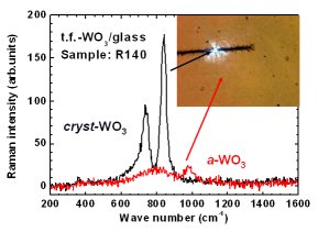
Confocal Spectromicroscopy of Amorphous and Nanocrystalline Tungsten Oxide Films.
(Presentation at the 10th International Conference on the Structure of Non-Crystalline Materials (NCM10), Praha, September 18-22, 2006.)
Introduction to Confocal Microscopy (for students).
LINKS
SOLinstruments - "Nanofinder S"
NT-MDT
TOKYO INSTRUMENTS,INC.
Symphotic Tii
Nikon Microscopy: Introduction to Confocal Microscopy
Nikon Objectives
Confocal Microscopy by Leica Microsystems
Olympus Confocal Microscopy
Images from the Microscope
VayTek, Inc. - digital imaging systems, deconvolution software, 3D volume visualization, etc.
Confocal Microscopy & Imaging at Purdue University.
Proscan. Special Instruments.
Edmund Optics.
Fluorescence Microscopy
Optical Components
KOZO Optics
OptiGrate
Qiooptiq
Chroma Technology Corp.
Kaiser Optical Systems
OmegaOptics
Semrock
Tydex
Iridian
Coherent
Exciton (Laser Dyes)
Horiba Jobin Yvon
Lasercomponents
Melles Griot
Plasma
Renishaw
Roithner Lasertechnik
Spectra Physics
CNI Laser
Klastech
Microlaser
Crystalaser
Integrated Optics
Andor
Acton Research
Nanonics
Raman Systems
Thorlabs
Allied Scientific Pro
OceanOptics
Linos
Standa
Eksmaoptics
Ekspla
WITec
Anda Optec (LV)
Z-Light (LV)
SPOT Imaging solutions
Digital Imaging Systems
Mesophotonics (SERS substrates)
Raman Spectra of Minerals
Raman spectra of minerals acquired at Caltech
Handbook of Minerals Raman Spectra
Rruff - an integrated database of Raman spectra, X-ray diffraction and chemistry data for minerals
Raman Spectroscopy
SpectroscopyNOW
Microscopy and Analysis
RRUFF database
The Internet Journal of Vibrational Spectroscopy
Journal of Raman Spectroscopy
ImageJ - image processing in Java.
Nikon's Small World - A stunning world seen through the microscope.
GALLERY
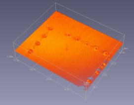
All images were taken by Dr. A. Kuzmin using the "Nanofinder-S" system, installed at ISSP (Riga, Latvia),
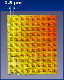
Calibrating Silicon Grating TGQ1 |
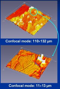
Confocal Imaging of CdWO4 Single-Crystal |
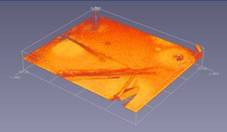
ZnO Needles on Silicon Substrate |
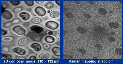
2D Chemical Phase Mapping in Oxide Films |
Materials for Optical Information Storage |
Structural Phase Transition
under Laser Irradiation |
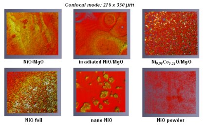
2D Confocal Imaging of Nickel Oxides |
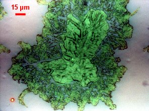
Dendrite-like Perrhenate Polycrystal |
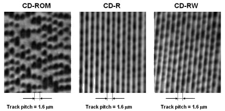
Optical Data Storage |
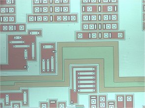
Electronic Chip |
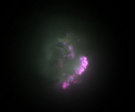
ZnO Luminescence
|
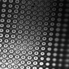
2D Grating
|
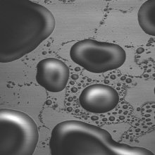
Liquid Bubbles
|
|